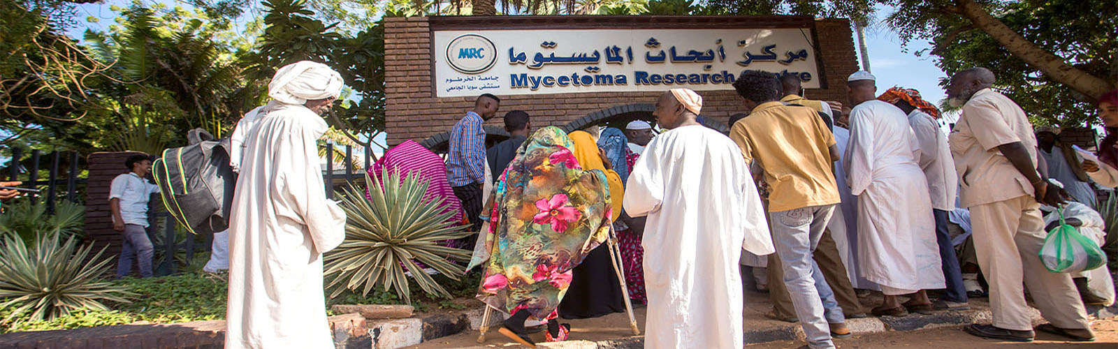
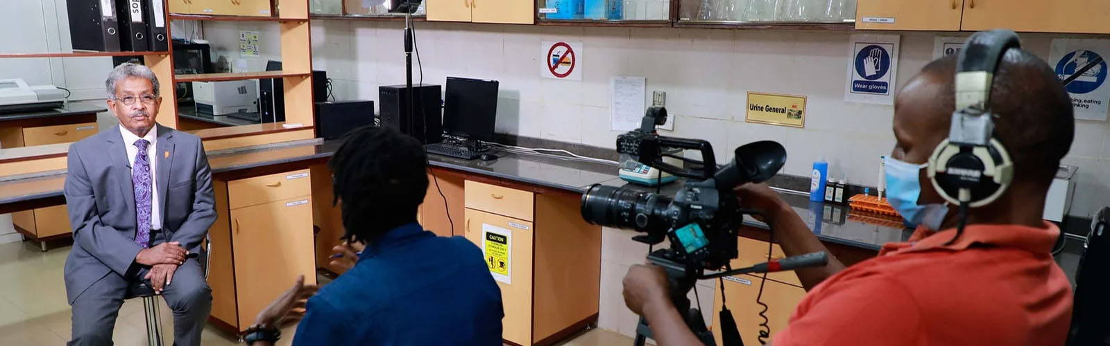
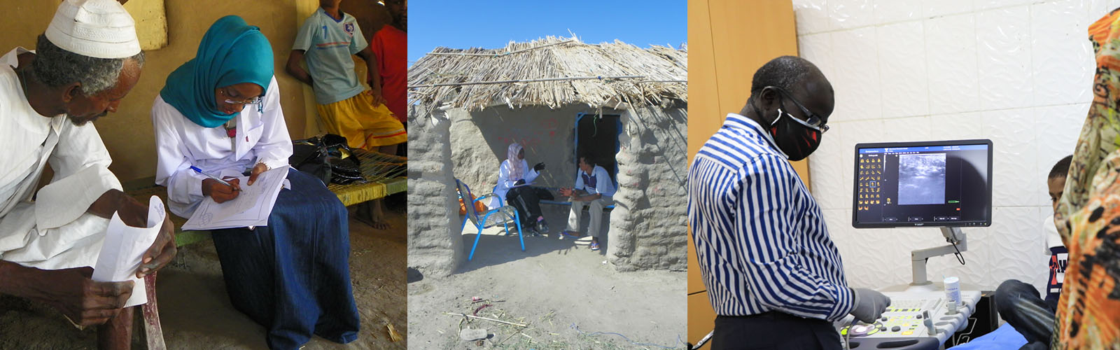
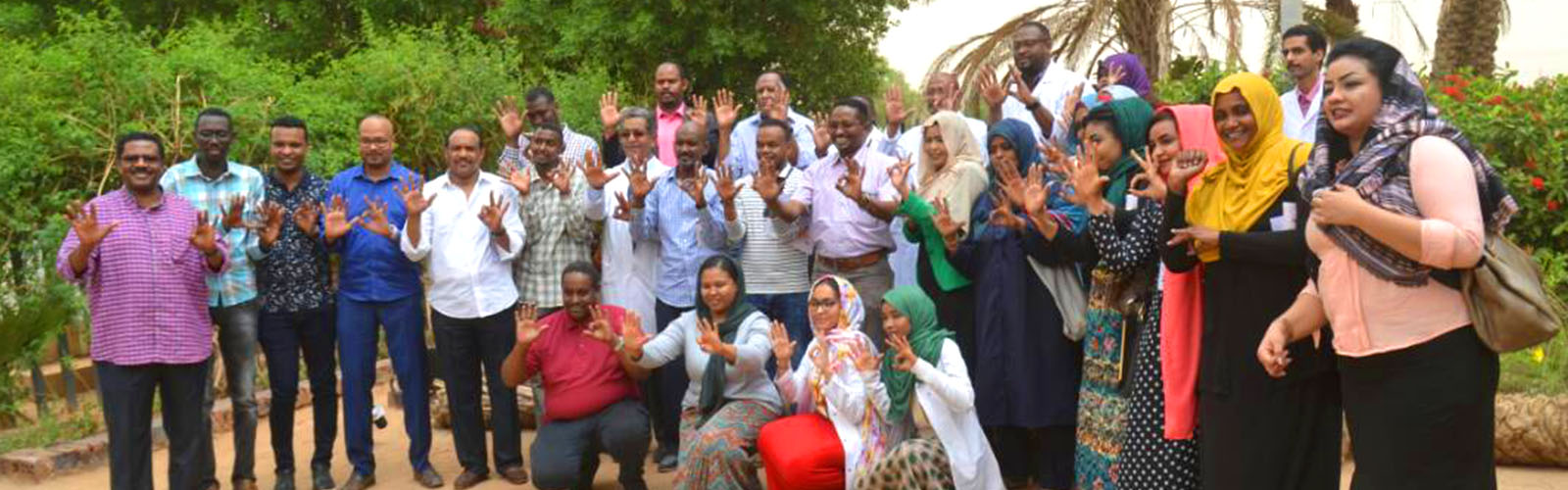
Magnetic Resonance Imaging

Magnetic Resonance Imaging (MRI) is useful for determining the extent of the lesions and the invasion of structures. It has greater sensitivity than radiographs, ultrasound, and CT.

MRI usually shows multiple 2-5 mm lesions of high signal intensity that indicates the granuloma, interspersed within a low-intensity matrix denoting the fibrous tissue. The ‘dot in circle’sign which indicates the presence of grains, is highly characteristic.

MRI

A MRI grading system; the Mycetoma Skin, Muscle, Bone Grading System (MSMBS), was recently proposed, it grades disease severity, compares patients and helps to manage them. The MRI appearance should be differentiated from chronic osteomyelitis, granulomas, soft tissue tumours, bone tuberculosis and cold abscesses. This procedure, however, is only possible in well-equipped hospitals.

In general, for small early lesions, ultrasound examination is recommended, meanwhile, radiography and MRI are recommended for large lesions.


banner