Medical Imaging Department

The Medical Imaging Department at the Mycetoma Research Center is devoted to providing top-notch medical care to individuals grappling with mycetoma. Our dedicated team of radiologists and staff members is committed to employing state-of-the-art imaging techniques to deliver cutting-edge services.
In our pursuit at the Mycetoma Research Center’s Medical Imaging Department, we aim to amalgamate expertise, advanced technology, and a dedication to education and research, contributing to the overall well-being of our mycetoma patients. Driven by a commitment to excellence, our department offers a comprehensive array of advanced imaging services tailored to the unique needs of mycetoma patients.
Under the leadership of Dr. Mustafa El Nour, our team utilises conventional X-ray examinations, ultrasound, MRI, and computed tomography scanning to ensure precise diagnoses and effective treatment planning. Additionally, we harness these sophisticated imaging techniques for educational purposes, incorporating them into teaching and research initiatives.
At the Medical Imaging Department, we strive to integrate expertise, advanced technology, and a commitment to education and research to enhance the overall well-being of our mycetoma patients.
Units
The department offers the patients conventional X-ray examination, ultrasound, MRI and computed tomography scanning, furthermore, these techniques are utilised for teaching, educational purposes, and research.
Meet the key members of our Medical Imaging Department:
Dr. Mustafa El Nour
Head of Department:
Consultant Radiologist and Assistant Professor of Radiology
Faculty of Medicine, University of Khartoum.
Mr. Omer El Hag
Senior Radiology Technologist
A skilled professional dedicated to supporting the department’s operations and contributing to the delivery of exceptional patient care.
Education and Training
The department offers several objective continuing professional development training and educational programmes for medical and health staff.
For more information, please contact info@mycetoma.edu.sd
Imaging Services
The service is available from Sunday to Thursday, 08:00-14:00
Request an appointment: info@mycetoma.edu.sd
Patient Care
The ultrasound unit usually performs more than 2,000 examinations each year using a state-of-the-art ultrasound machine. A portable ultrasound machine is available for field epidemiological surveys for early detection and surveillance in mycetoma endemic regions and patient care at the mycetoma satellite units. Ultrasound-guided biopsies and Fine needle aspiration cytology procedures are conducted routinely for deep-seated lesions diagnosis.
Ultrasonic Imaging of Mycetoma
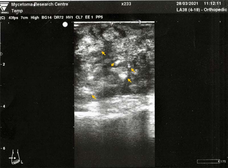
The mycetoma grains, capsule, and the accompanying inflammatory granuloma have characteristic ultrasonic appearances. Ultrasound imaging can differentiate between eumycetoma, actinomycetoma, mycetoma and other non-mycetoma lesions. The grains produce numerous sharp, bright hyperreflective echoes consistent with the black grains in eumycetoma lesions. The grain cement substance is most probably the origin of these sharp echoes. Also, there are multiple thick-walled cavities with absent acoustic enhancement. In actinomycetoma lesions, the findings are similar, but the grains are less distinct. This may be due to their smaller size and consistency, the individual embedding of the grains or the absence of the cement substances in a few.
The ultrasonic diagnosis of mycetoma is more precise and accurate in lesions with no sinuses. The size and extent of the lesion can be accurately determined ultrasonically, which is useful in planning surgical incisions and procedures.
Fahal AH, Sheikh HE, EL Lider MA, Homeida MA, EL Arabi YE, Mahgoub ES, Ultrasonic imaging in mycetoma. BriJSurg. 1997; 78: 765-766.
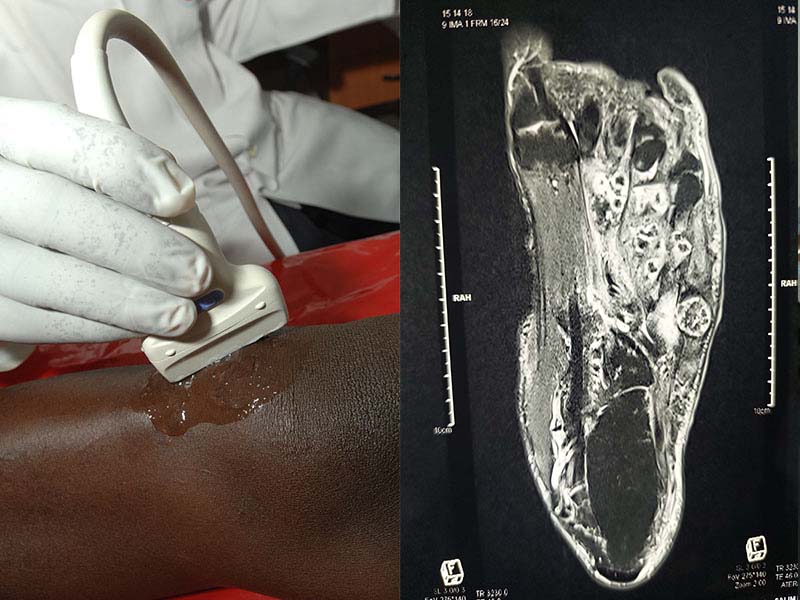
MRI Scanning
MRI is non-invasive imaging, and it can characterise the soft tissue masses of mycetoma and aid in early diagnosis. The mycetoma lesion in the MRI appears in the form of the dot-in-circle sign, which is a unique pathological feature of mycetoma and it is a highly specific sign for mycetoma. In advanced cases, MRI is essential to plan treatment options.
EL Shamy, ME, Fahal AH, Shakir MY, Homedia MMA. New MRI Grading System for the Diagnosis and Management of Mycetoma. Trans R Soc Trop Med Hyg. 2012; 106(12):738-42.