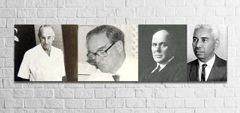
Mycetoma Pioneers

Historical Footage & Pioneers of Mycetoma in the Sudan
Written by Dr.Tarik Elhadd, Consultant Physician.
Sudan seems to be the mycetoma homeland. The disease is known in the Sudan, before the advent of modern medicine, by its present common name of “Nebit” meaning growth. During the last years of the era of the Turco-Egyptian Sudan (1821-1885), the senior doctor in Khartoum Hospital was Dr. Nessib Selim, an Egyptian doctor, who was noted to be a physician and a surgeon of repute. Dr. Selim used to operate on cases of Madura foot and bladder stones. Treatment by cautery and/or amputation was practiced by native doctors since the time of the Mahdiyya (1885-1898).
Dr. Andrew Balfouer, a Scottish physician and the first director of the Wellcome Tropical Research Laboratory in Khartoum (1903-1913) was the first to report on a case of mycetoma in the Sudan in 1904. Dr. Balfour noted that the disease was common amongst Northern Sudanese, that the foot was affected most and that the commonest type was the black grain variety.
The first case from Southern Sudan was reported in 1908 from Borr, in the Upper Nile Province by Dr. Chris Morley Wenyon, the eminent London microbiologist when he was the Travelling Pathologist of the Sudan government.
Extensive studies of two causal organisms were carried out by Dr. Albert Chalmers, Director of the Wellcome Tropical Research Laboratories (1913-1920), together with Captain R.G. Archibald, pathologist, and Dr. J.B. Christopherson, Director of Khartoum and Omdurman Civil Hospitals.They carried out valuable systemic mycological and pathological studies of the causal organisms. Chalmers and Archibald gave quite an elaborative and specific definition of mycetoma. For the first time, they introduced the terms Maduromycoses and Actinomycoses, proceeded afterwards to classify the mycetomas into Maduromycetoma the grains of which are composed of large segmented mycelia and Actinomycetoma with grains composed of fine non-segmented filaments.
In 1931, Grantham-Hill, the senior surgeon, Khartoum Hospital made a detailed clinical study of 184 cases out of which 64% were of the black variety and 36% were yellow. Noting that the yellow type is actinomycotic and the black type is maduromycotic, he discussed the relative virulence of the two types. He thought that the actinomycotic mycetoma was more virulent, infiltrates gradually and once it penetrates the periosteum it disseminates rapidly in the bone while the black maduromycotic mycetoma forms a usually localised subcutaneous tumour. He doubted the value of medical treatment by various drugs suggested up to that date. He thought that the best routine treatment was surgical and that the key to success lies in early recognition and complete removal. In the case of black maduromycosis, the pseudo-capsule can readily be identified by its bluish colour and dissection follows its outer surface. In the absence of sinuses, an incision is made into the tumour to identify its nature. If it is found to be mycetoma, fresh instruments are taken and a circumscribing incision made at a distance at about one centimetre from the apparent margin of the growth. This caution in surgical removal reflects an excellent knowledge of the high rate of recurrence in the case of inadequate removal.
Grantham-Hill was aware that amputations would deter patients from attending to thehospital and therefore encouraged early detection. He, therefore, defended local removal as much as possible. In his series of 184 cases, 141 (77%) were treated by local removal and 43 (23%) by amputation. He gave support to the common belief that the “Nebit” usually follows a thorn prick by saying that in 30% of his patients with mycetoma of lessthan six months duration, thorns were actually found embedded in the growth after removal at operation.
Dr. Robert Kirk of the Sudan Medical Service, jointly with Dr. Aldridge were the first to report a case of mycetoma in the eyelid. Dr. Kirk later became the first Professor of Pathology at theUniversity of Khartoum.
Peter Abbott, a physician & surgeon at Wad Medani Civil Hospital, was awarded the degree of M.D. (1954) from Cambridge University on his Clinical and Epidemiological Studies of Mycetoma. He also addressed the Royal Society of Tropical Medicine and Hygiene in London in December 1956 on mycetoma in Sudan. Abbott sent specimens from his cases abroad to Dr. Walker’s Mycological Reference Laboratory at the London School of Hygiene and Tropical Medicine and to Professor Juan E. MacKinnon in Montevideo in Uruguay for mycological identification.
Abbott carried out in vitro trials with antibiotics against Madurella mycetomiand Streptomyces somaliensis. He found that the growth of M. mycetomiwas unaffected by chloramphenicol, oxytetracycline, carbomycin and polymyxin B. S. somaliensis was markedly sensitive to all these antibiotics except Polymyxin B.
Regarding surgical treatment, Abbott reaffirmed the significance of either removal of the tumour in toto or if its border was ill-defined, a margin of healthy tissue was removed as well to guard against recurrence. Abbott brought to light the fact that mycetoma in the Sudan was a serious and common disease leading to the loss by amputation of many limbs. He reported on 1231 cases of mycetoma who were admitted to hospitals in Sudan or were seen in outpatient clinics in two and half years. His own study was based on 207 cases and therefore was referred to as the largest single study on mycetoma for many years. From the specimens of Abbott, Professor MacKinnon settled the identity of black grain mycetoma in Sudan as M. mycetomi and showed that it is not different to merit the Glenospora khartoumensis coined early by Chalmers and Archibald 1916 and that the yellow type is due to S. somaliensis.
Abbott noted that trophic and neurological changes were absent in mycetoma and on dissection of some of the mycetomata, glistening white tendons and nerves lay unaffected in the middle of damaged tissue.
In 1964, Professor James. B. Lynch, Professor of Pathology, Faculty of Medicine, University of Khartoum gave a Hunterian lecture on “Mycetoma in Sudan” to the Royal College of Surgeons of England. He reiterated the findings on the epidemiology of the diseases and gave a nice study of its pathology. He studied both the causal organisms and the tissue reaction. He estimated the incidence of mycetoma in the Sudan to vary from 300-400 new hospital cases annually; this figure does not take into account the considerable number of outpatients and those seen at peripheral dispensaries where qualified doctors might not even be present. However, recent estimates on the incidence of mycetoma have not been reported.
Mr. Ibrahim Moghraby (1967) who worked for many years as a surgeon at Wad Medani Hospital, centre of endemic area for mycetoma, presented to the 6th Arab Medical Conference in Khartoum a study on “Mycetoma in the Gezeria, a public health problem”.
In the early seventies of the last century, the Sudan responded to the Mycetoma threat by establishing The Khartoum North Mycetoma Clinic and ward for patients’ medical care and research.
It was established through the help of the World Health Organization, the Ministry of Overseas Development of the United Kingdom and the idea was initiated by the late Professor Julian Taylor, Professor of Surgery, University of Khartoum, the clinic was supported by the Ministry of Health of the Sudan and the University of Khartoum.
The mycetoma clinic and mycetoma ward were established at the Khartoum North Civil Hospital. The objective of this project was to conduct clinical trials for thetreatment of mycetoma and take full care of the patients.However, the availability of patients and technical capabilities of the team in charge made it possible to carry out further research on serological diagnosis.
