Histopathology of Mycetoma
The Histopathology Department at the MRC provides extensive assistance in diagnosing mycetoma through histopathological examination of surgical biopsies, cell block specimens, and immunohistochemical analysis of specific samples. Additionally, the department is responsible for preparing pathology museum specimens.
Biological Material Management
The histopathological diagnosis of mycetoma involves the collection of adequate surgical biopsies or aspirated cell blocks. This involves the careful collection, handling, and storage of various biological materials according to Standard Operating Procedures (SOPs):
The Surgical biopsy:
A surgical biopsy can be procured through wide local surgical excision, incision biopsies, or tru-cut needle biopsy. It is recommended that the biopsy be conducted under general, spinal, or regional anaesthesia. The use of local anaesthesia should be avoided due to the superficial and potentially inadequate nature of the specimen, as well as the associated discomfort.
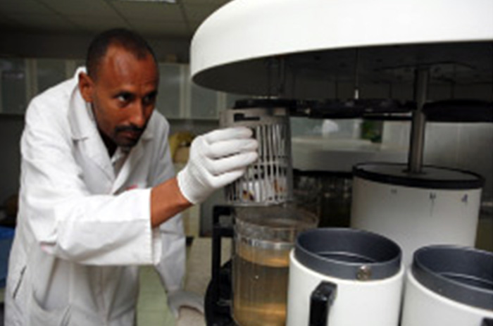
The surgical biopsies should be handled immediately by the laboratory technologist at the surgical theatre; they should be divided into two parts: one part for the grain culture and PCR identification, which should be placed in normal saline and another part for histopathological examinations. The later part should be fixed immediately in a 10% formal saline solution.
The material should be adequate in amount, free of contaminants, collected in proper containers, and transported speedily to the laboratory for the collection sites. Each sample should be labeled clearly with patient information, date, and sample type to avoid any mix-ups. Samples should be transported to the laboratory in a timely manner, maintaining appropriate temperature conditions to prevent degradation.
The biopsies should be processed for the preparation of tissue blocks and histopathological examination.
Mycetoma causative microorganism microscopical appearance
Host tissue reactions appearance.

Staff
- Dr Badreldin M. Yousif
- Dr Nadia EL Dawi
- Miss Omnia Babiker
Publications List:
- Fahal AH, el Toum EA, el Hassan AM, Mahgoub ES, Gumaa SA. The host tissue reaction to Madurella mycetomatis: new classification. J Med Vet Mycol. 1995 Jan-Feb;33(1):15-7. PMID: 7650573.
- Ahmed AA, van de Sande W, Fahal AH. Mycetoma laboratory diagnosis: Review article. PLoS Negl Trop Dis. 2017 Aug 24;11(8):e0005638. doi: 10.1371/journal.pntd.0005638. PMID: 28837657; PMCID: PMC5570215.
- el Hassan AM, Fahal AH, Ahmed AO, Ismail A, Veress B. The immunopathology of actinomycetoma lesions caused by Streptomyces somaliensis. Trans R Soc Trop Med Hyg. 2001 Jan-Feb;95(1):89-92. doi: 10.1016/s0035-9203(01)90346-3. PMID: 11280076.
- Fahal AH, el Toum EA, el Hassan AM, Mahgoub ES, Gumaa SA. A preliminary study on the ultrastructure of Actinomadura pelletieri and its host tissue reaction. J Med Vet Mycol. 1994;32(5):343-8. PMID: 7844700.